Discectomy - Microdiscectomy
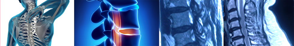
An intervertebral disc is a kidney-shaped structure located between each pair of vertebrae. They are designed to redistribute forces incurred by the spinal column when sitting, standing or lifting.
The intervertebral disc is composed of two types of cartilage. There is an inner semi-liquid cartilage (nucleus pulposus) surrounded by several layers of fibrous rings of cartilage (annulus fibrosis). Together they form a self-contained unit.
The intervertebral disc is strong and stable. When an object is lifted, the force is transmitted directly onto the semi-liquid center and then redistributed radially to the outer fibrous rings. As the force is redistributed, the rings resist deformation. It is this combination that enables the disc to act like a shock absorber.
Etiology of disc rupture or herniation
Repetitive bending and twisting or trauma to the back can create a shear stress across the disc. Over time this stress may cause the outer fibrous rings to break down, one layer at a time. Gradually, the semi-liquid center of the disc will work its way through the outer ring and push on a nerve. This is known as a ruptured, slipped or herniated disc.
There are five discs in your lower back, located between each pair of vertebrae from L1 to S1. The two lowest discs, L4-5 and L5-S1 most commonly rupture. Pressure on a nerve may cause pain, numbness, weakness, tingling, or loss of a reflex. The distribution of symptoms varies depending on which nerve root is involved.
The center of the disc protrudes through the outer ring (annulus) and subsequently puts pressure on a nerve, causing pain to radiate down the patient’s leg and into the foot.
Micro discectomy
Microdiscectomy is a minimally invasive surgical procedure in which a portion of a herniated nucleus pulpolsus is removed by way of a surgical instrument or laser while using an operating microscope for magnification.
Microdiscectomy uses a special microscope that lets the surgeon view the damaged disc and compressed nerves. This enables them to make more precise incisions, with less corresponding damage to neighboring tissue it is important to realize that a microdiscectomy is quite effective in relieving pain in the buttocks or pain that travels down the leg; however, it is not particularly effective in relieving back pain. Back pain is typically treated with aggressive rehabilitation. The primary reason to proceed with surgery is to relieve pain in the buttocks or leg sooner than would be accomplished without surgery.
The compression on the nerve root can cause substantial leg pain, and while it may take weeks or months for the nerve root to fully heal and for any numbness or weakness to get better, patients normally feel relief from leg pain almost immediately after a microdiscectomy surgery.
Microdiscectomy enables surgeons to treat a number of spinal disorders, such as :
- Degenerative disc
- Herniated disc
- Spinal fractures
- Spinal tumors
- Infections
- Instability
Benefits of Microdiscectomy
- Smaller incisions
- Reduced blood loss
- Less pain
- Smaller scars
- Reduced muscle retraction
- Shorter hospital stay
- Quicker recovery
- Faster return to normal activities
It is intended to decrease pain and restore normal movement and function in the patient, and is considered for patients who meet the following criteria :
- Severe leg pain, numbness, and/or weakness that prevents normal functioning
- Patients who have experienced leg pain for four to six weeks and who have tried conservative treatment (such as oral steroids, epidural steroid injections, NSAID’s, and physical therapy) without successfully relieving the pain.
- However, it is not advisable to wait too long before having this surgery, because the results are not as good if the surgery is postponed more than three to six months. Besides time, one needs to also factor in the level of the pain and the amount of disability the patient is experiencing. If the symptoms are mild, a longer course of conservative treatment may be reasonable, whereas if the symptoms are severe more immediate surgery is reasonable. Failure to improve after at least four weeks of nonsurgical treatment.
- Physical examination indicates that improvement is likely after surgery
- Patient has cauda equina syndrome, requiring emergency intervention to alleviate pain, which has not improved within a reasonable time period.
Microdiscectomy is performed through a small (1 inch to 1 1/2 inch) incision in the midline of the back.
-
- First, the back muscles (erector spinae) are lifted off the bony arch (lamina) of the spine. Since these back muscles run vertically, they can be moved out of the way rather than cut.
- The surgeon is then able to enter the spine by removing a membrane over the nerve roots (ligamentum flavum), and uses either operating glasses or an operating microscope to visualize the nerve root.
- Often, a small portion of the inside facet joint is removed both to facilitate access to the nerve root and to relieve pressure over the nerve.
- The nerve root is then moved to the side and the disc material is removed from under the nerve root.
Decompression & Fixation
Introduction
The lower back, or lumbar region, is made up of the five lowest vertebrae (L1 – L5) that are just above the base of the spine. The lumbar region supports the bulk of the weight of the upper body and is the most common area of back pain.
Decompression is a surgical procedure that is performed to alleviate pain caused by pinched nerves (neural impingement). Compression of the nerve roots and narrowing of the lumbar spinal canal can be caused by the intervertebral disc, ligaments and overgrowth of bone (osteophytes). Compression of the nerve roots can lead to pain in the legs (calves) on walking, numbness and weakness in the legs on walking and occasionally bowel and bladder complaints.
Candidate for decompression if you have :
- Significant pain, weakness, or numbness in your leg or foot
- Leg pain worse than back pain
- Not improved with physical therapy or medication
- Difficulty walking or standing that affects your quality of life
- Diagnostic tests (MRI, CT, myelogram) that show stenosis in the central canal or lateral recess.
- Spinal stenosis, a herniated disc
- Isthmic or degenerative spondylolisthesis
- A spinal tumor
Spinal decompression can be performed anywhere along the spine from the neck (cervical) to the lower back (lumbar). The procedure is performed through a surgical incision in the back (posterior).The lamina is the bone that forms the backside of the spinal canal and makes a roof over the spinal cord. Removing the lamina and other soft tissues gives more room for the nerves and allows for removal of bone spurs. Depending on the extent of stenosis, one vertebra (single-level) or more (multi-level) may be involved.
There are several types of decompression surgery:
Laminectomy is the removal of the entire bony lamina, a portion of the enlarged facet joints, and the thickened ligaments overlying the spinal cord and nerves. Laminotomy is the removal of a small portion of the lamina and ligaments, usually on one side. Foraminotomy is the removal of bone around the neural foramen – the space between vertebrae where the nerve root exits the spinal canal. This method is used when disc degeneration has caused the height of the foramen to collapse, resulting in a pincheced nerve. It can be performed with a laminectomy or laminotomy. Laminoplasty is the expansion of the spinal canal by cutting the laminae on one side and swinging them open like a door. It is used only in the cervical area.
Spinal fusion may be done at the same time to help stabilize sections of the spine treated with laminectomy or laminectomies at multiple vertebrae levels. Fusion is the joining of two vertebrae with a bone graft held together with hardware such as plates, rods, hooks, pedicle screws, or cages. The goal of the bone graft is to join the vertebrae above and below to form one solid piece of bone. There are several ways to create a fusion.
Advantage of – Fusing the joint
- Prevents the spinal stenosis from recurring and can help eliminate pain from an unstable spine
- Prevent movement between the involved vertebrae and realign the spinal column so as to reduce the pain. The screws are rods are made of either titanium or stainless steel and are well tolerated by the body.
Vertebroplasty
Vertebroplasty is a minimally invasive, day care procedure used to treat vertebral compression fractures caused by painful osteoporosis and metastatic tumors. During vertebroplasty, A local anesthetic is used to numb the affected area of the patient’s spine, where the surgeon inserts one or two needles guidance through a small incision in the patient’s skin. Under under C- arm, the surgeon inserts the needles into the fractured vertebra through pedicals and slowly injects a small amount of bone cement into the vertebra at fracture site. The bone cement hardens quickly. The patient is kept for observation for a few hours following the procedure. In rare cases, the patient is kept overnight for observation.
Vertebroplasty can prevent further collapse of the vertebra, height loss and spine curvature.
For further details visit “FAQ FOR VERTEBROPLASTY”