Knee Replacement
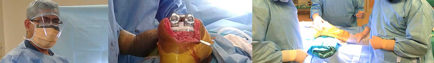
Total Knee Replacement Surgery
Introduction
Knee replacement surgery was first performed in 1968. Since then, improvements in surgical materials and techniques have greatly increased its effectiveness. Total knee replacements are one of the most successful procedures in Orthopedics
Knee Joint
The knee is the largest joint in the body and having healthy knees is required to perform most everyday activities. The knee is made up of the lower end of the thighbone (femur), the upper end of the shinbone (tibia), and the kneecap (patella). The ends of these three bones where they touch are covered with articular cartilage, a smooth substance that protects the bones and enables them to move easily.
Details of Surgery
Knee replacement (also called knee arthroplasty) might be more accurately termed a knee “resurfacing” because only the surface of the bones are actually replaced. There are four basic steps to a knee replacement procedure.
There are four basic steps to a knee replacement procedure
- Prepare the bone: The damaged cartilage surfaces at the ends of the femur and tibia are removed along with a small amount of underlying bone.
- Position the metal implants: The removed cartilage and bone is replaced with metal components that recreate the surface of the joint. These metal parts may be cemented or “press-fit” into the bone.
- Resurface the patella: The undersurface of the patella (kneecap) is cut and resurfaced with a plastic button. Some surgeons do not resurface the patella, depending upon the case.
- Insert a spacer: A medical-grade plastic spacer is inserted between the metal components to create a smooth gliding surface.
- Deformity correction: During the operation any deformities must be corrected, and the ligaments balanced so that the knee has a good range of movement and is stable and aligned.
Total Knee Implant
- Femoral Component: In a knee implant, the femoral component, made of metal, curves up arounds the end of the femur (or thighbone). It has a central groove allowing the patella (or kneecap) to move up and down smoothly as the knee joint bends and straightens.
- Tibial Component: The tibial component of a knee implant is a flat metal platform with a polyethylene insert or spacer. These have a double dish configuration for the femoral condyles and also either a notch to accommodate the cruciate ligaments (cruciate sparing) or a cam structure to take their place (cruciate sacrificing).
- Patellar Componen: The patellar ‘button’ is a dome-shaped piece of ultrahigh molecular weight polyethylene that replicates the surface of the kneecap.
Unconstrained :- Cruciate substituting/ Cruciate retaining
- Fixed Bearing: The polyethylene cushion of the tibial component is fixed to the metal platform base.
- Mobile Bearing: The difference between a fixed-bearing implant and a mobile bearing implant is in the bearing surface. They allow patients a few degrees of greater rotation to the medial and lateral sides of their knee.
- Medial Pivot (also known as Rotating Platform): In a rotating platform, the polyethylene insert can rotate slightly around a conical post, thereby copying the activity of the natural knee joint.
- Posterior Cruciate Ligament (PCL)-Retaining: Another important aspect of a total knee replacement is the treatment of the Posterior Cruciate Ligament which prevents the femur from shunting back on the top of the tibia when it is flexed – sometimes referred to as ‘roll back’. Depending upon its condition, this ligament can be kept (retained) or sacrified.
Commonly available / used implant brands
- Stryker – Knee Systms – Scorpio NRG, Scorpio Single Axis
- Depuy – PFC Sigma, RPF
- Smith Nephew – Genesis II, Journey, Technology
- VERILAST-OXINIUM- alloy and a highly cross-linked polyethylene(XLPE)
- VISIONAIRE – Patient Matched Instrumentation-uses the patient’s own MRI and full leg X-Ray to design cutting blocks specific to that patient.
UKR-Partial knee replacement
The knee is generally divided into three “compartments”: medial (the inside part of the knee), lateral (the outside), and patellofemoral (the joint between the kneecap and the thighbone). patients having arthritis of all three compartments need total knee replacement .patients with wear confined primarily to one compartment, usually the medial, and may be candidates for unicompartmental knee replacement or High TIbial Osteotomy. Advantages of UKA compared to TKA include smaller incision, easier post-op rehabilitation, better post-operative range of motion, shorter hospital stay, less blood loss, lower risk of infection, stiffness, and blood clots.
Hip Replacement
Anatomy
Hip is one of the body’s largest joints. It is a ball-and-socket joint. The socket is formed by the acetabulum, which is part of the large pelvis bone. The ball is the femoral head, which is the upper end of the femur (thighbone).The bone surfaces of the ball and socket are covered with articular cartilage, a smooth tissue that cushions the ends of the bones and enables them to move easily.
A thin tissue called synovial membrane surrounds the hip joint. In a healthy hip, this membrane makes a small amount of fluid that lubricates the cartilage and eliminates almost all friction during hip movement. Bands of tissue called ligaments (the hip capsule) connect the ball to the socket and provide stability to the joint.
Total Hip Replacement
Total hip replacement surgery is a major orthopedic procedure which is designed to relieve pain by the removal of a damaged hip joint and the replacement of that joint with prosthesis and help the patient return to everyday activities. Traditional and minimally invasive hip surgeries are both options for those suffering from pain or immobility of the hip joints.
Description
In a total hip replacement (also called total hip arthroplasty), the damaged bone and cartilage is removed and replaced with prosthetic components.
- The damaged femoral head is removed and replaced with a metal stem that is placed into the hollow center of the femur. The femoral stem may be either cemented or “press fit” into the bone.
- A metal or ceramic ball is placed on the upper part of the stem. This ball replaces the damaged femoral head that was removed.
- The damaged cartilage surface of the socket (acetabulum) is removed and replaced with a metal socket. Screws or cement are sometimes used to hold the socket in place.
- A plastic, ceramic, or metal spacer is inserted between the new ball and the socket to allow for a smooth gliding surface.
Anesthesia
After admission, patient will be evaluated by a member of the anesthesia team. The most common types of anesthesia are general anesthesia (you are put to sleep) or spinal, epidural, or regional nerve block anesthesia (you are awake but your body is numb from the waist down). The anesthesia team, with your input, will determine which type of anesthesia will be best for you.
Implant Components
Many different types of designs and materials are currently used in artificial hip joints. All of them consist of two basic components: the ball component (made of highly polished strong metal or ceramic material) and the socket component (a durable cup of plastic, ceramic or metal, which may have an outer metal shell).
The prosthetic components may be “press fit” into the bone to allow your bone to grow onto the components or they may be cemented into place. The decision to press fit or to cement the components is based on a number of factors, such as the quality and strength of your bone. A combination of a cemented stem and a non-cemented socket may also be used.
Your orthopaedic surgeon will choose the type of prosthesis that best meets your needs.
Dr Punit take a comprehensive approach to the treatment of hip pain including non-surgical therapies and total hip replacements. Whether you are simply in need of physical therapy or total hip replacement surgery. Dr. Punit is always with you to help you through every step of the diagnosis, treatment and rehabilitation process. If you are in need of treatment for hip pain and/or hip replacement surgery, schedule a consultation with Dr Punit today.
Anatomy
Shoulder is made up of three bones, upper arm bone (humerus), shoulder blade (scapula), and collarbone (clavicle). The shoulder is a ball-and-socket joint: The ball, or head, of your upper arm bone fits into a shallow socket in your shoulder blade. This socket is called the glenoid. The surfaces of the bones where they touch are covered with articular cartilage, a smooth substance that protects the bones and enables them to move easily. A thin, smooth tissue called synovial membrane covers all remaining surfaces inside the shoulder joint.The muscles and tendons that surround the shoulder provide stability and support.Conditions for shoulder replacement
- Osteoarthritis (Degenerative Joint Disease)This is an age-related “wear and tear” type of arthritis. It usually occurs in people 50 years of age and older, but may occur in younger people, too. The cartilage that cushions the bones of the shoulder softens and wears away. The bones then rub against one another. Over time, the shoulder joint slowly becomes stiff and painful
- Rheumatoid Arthritis : This is a disease in which the synovial membrane that surrounds the joint becomes inflamed and thickened. This chronic inflammation can damage the cartilage and eventually cause cartilage loss, pain, and stiffness. Rheumatoid arthritis is the most common form of a group of disorders termed “inflammatory arthritis.”
- Post-traumatic Arthritis: This can follow a serious shoulder injury. Fractures of the bones that make up the shoulder or tears of the shoulder tendons or ligaments may damage the articular cartilage over time. This causes shoulder pain and limits shoulder function.
- Rotator Cuff Tear Arthropathy A patient with a very large, long-standing rotator cuff tear may develop cuff tear arthropathy. In this condition, the changes in the shoulder joint due to the rotator cuff tear may lead to arthritis and destruction of the joint cartilage.
- Avascular Necrosis (Osteonecrosis): Avascular necrosis is a painful condition that occurs when the blood supply to the bone is disrupted. Because bone cells die without a blood supply, osteonecrosis can ultimately cause destruction of the shoulder joint and lead to arthritis.
- Severe Fractures : A severe fracture of the shoulder is another common reason people have shoulder replacements. When the head of the upper arm bone is shattered, it may be very difficult to put the pieces of bone back in place.
- Failed Previous Shoulder Replacement Surgery:-Although uncommon, some shoulder replacements fail, most often because of implant loosening, wear, infection, and dislocation. When this occurs, a second joint replacement surgery — called a revision surgery — may be necessary.
Indications for Shoulder Joint Replacement
People who benefit from surgery often have :- Severe shoulder pain that interferes with everyday activities, such as, dressing, toileting, and washing.
- Moderate to severe pain while resting. This pain may be severe enough to prevent a good night’s sleep.
- Loss of motion and/or weakness in the shoulder.
- Failure to substantially improve with other treatments such as anti-inflammatory medications, cortisone injections, or physical therapy.
- X-rays or MRI shows arthritis or other problems.
Total shoulder replacement implant
The humeral implant consists of a metal ball that replaces the head of the humerus, and a stem that is secured into the main arm bone (humerus).The humeral stem is made of titanium for maximum strength. The head is made of cobalt chrome to provide a smooth surface for movement with the glenoid component, which is made of polyethylene. The metal ball and stem are selected by the surgeon from multiple sizes to fit the contour and shape of each patient’s humerus. This two-piece construction is known as a modular implant. This modularity allows surgeons to closely replicate the natural shoulder. If the surgeon uses only the metal humeral components (humeral head and stem), the procedure is called a partial shoulder replacement. If the surgeon uses both the humeral components and the glenoid implant, the procedure is called a total shoulder replacement. Surgeons will decide which procedure to use based on the extent of damage to their patients’ shoulders.Shoulder Replacement Options
Shoulder replacement surgery is highly technical. It should be performed by a surgical team with experience in this procedure.Total Shoulder Replacement
The typical total shoulder replacement involves replacing the arthritic joint surfaces with a highly polished metal ball attached to a stem, and plastic socket.t They may be either cemented or “press fit” into the bone. If the bone is of good quality, surgeon may choose to use a non-cemented (press-fit) humeral component. If the bone is soft, the humeral component may be implanted with bone cement. In most cases, an all-plastic glenoid (socket) component is implanted with bone cement.Implantation of a glenoid component is not advised ift
- The glenoid has good cartilage
- The glenoid bone is severely deficient
- The rotator cuff tendons are irreparably torn
Shoulder Hemiarthroplasty
Depending on the condition of your shoulder, surgeon may replace only the ball. This procedure is called a hemiarthroplasty. Indications for a hemiarthroplasty include:- Arthritis that only involves the head of the humerus with a glenoid that has a healthy and intact cartilage surface
- Shoulders with severely weakened bone in the glenoid
- Some shoulders with severely torn rotator cuff tendons and arthritis the humeral head is severely fractured but the socket is normal.
Resurfacing Hemiarthroplasty
Resurfacing hemiarthroplasty involves replacing just the joint surface of the humeral head with a cap-like prosthesis without a stem. With its bone preserving advantage, it offers those with arthritis of the shoulder an alternative to the standard shoulder replacement. Resurfacing hemiarthroplasty may be an option for you if :Anatomy
The ankle joint is made up of three bones: the lower end of the tibia (shinbone), the fibula (the small bone of the lower leg), and the talus, the bone that fits into the socket formed by the tibia and fibula. The talus sits on top of the calcaneus (the heel bone). The talus moves mainly in one direction. It works like a hinge to allow your foot to move up and down.
Ligaments on both sides of the ankle joint help hold the bones together. Many tendons cross the ankle to move the ankle and the toes. (Ligaments connect bone to bone, while tendons connect muscle to bone.) The large Achilles tendon at the back of the ankle is the most powerful tendon in the foot. It connects the calf muscles to the heel bone and gives the foot the power for walking, running, and jumping.
Inside the joint, the bones are covered with a slick material called articular cartilage. Articular cartilage is the material that allows the bones to move smoothly against one another in the joints of the body. The cartilage lining is about one-quarter of an inch thick in most joints that carry body weight, such as the ankle, hip, or knee. It is soft enough to allow for shock absorption but tough enough to last a lifetime, as long as it is not injured.
Introduction
Surgery to replace the ankle joint with an artificial joint (called ankle arthroplasty) is becoming more common. Recent advances in the design of the artificial ankle and changes in the way the operation is performed have made artificial ankle replacement a growing alternative to ankle fusion for the treatment of ankle arthritis. The restoration of range of motion is the key feature in favor of ankle replacement with respect to arthrodesis. There is another very important aspect to ankle replacement in that it avoids the stresses that occur following ankle fusion or arthrodesis. When an ankle joint is fused, there is of course no up and down movement in the ankle. There does however remain for some patients a limited amount of up and down movement which occur in the adjacent joints.
Indications :The ideal patient, is someone who is over the age of 50, is not too heavy, and is not extremely active